
Of all the numbers in skin-cancer medicine, one contrast matters more than any other. When melanoma is found while it is still confined to the skin, survival is very high. The five-year relative survival rate for melanoma is around 95% overall, and it approaches nearly 100% when the cancer is caught at a localized stage before it has spread. Once it reaches distant organs, the outlook falls sharply, to an average of roughly 17% at the most advanced stage. The disease itself does not change on that journey. Only the timing of when someone noticed it does. That is why early detection is not a marketing phrase in dermatology. It is the entire game.
This article looks at what the evidence actually says about finding melanoma sooner, why the human eye alone is not enough, and what both patients and clinicians can do to shift the odd in their favour.
The biology of “too late”
Melanoma is graded largely by how deeply it has grown into the skin, a measurement called Breslow thickness. Thinner cancers are less likely to spread, and prognosis is strongly tied to that thickness. Thin, early lesions are usually cured with a simple excision performed under local anaesthetic. As a tumour grows downward, it gains access to blood vessels and the lymphatic system, and the chance of it seeding elsewhere in the body climbs steeply.
This is why the field is so preoccupied with time. The same melanoma found a few months earlier can be, in prognostic terms, a genuinely different disease. It can end with a small scar rather than years of treatment. Nothing else in the melanoma story carries this much leverage.
The limits of looking
It is tempting to assume that a dangerous spot looks obviously wrong. In reality, early melanomas are often subtle, and the nakeds eye is a surprisingly blunts instrument. The accuracy of diagnosing cutaneous melanoma with the unaided eye is only about 60%. That is not a criticism of anyone’s eyesight. It is a limit of what surface-level looking can reveal about processes happening beneath the skin.
A useful starting framework for the public is the ABCDE rule, developed to flag which lesions are worth showing a professional:
- Asymmetry: one half does not match the other.
- Border: edges that are irregular, notched, or blurred.
- Colour: more than one shade, or uneven distribution across the spot.
- Diameter: larger than about 6 mm (a pencil eraser), though melanomas can be smaller.
- Evolving: any change in size, shape, colour, or sensation over time.
The ABCDE rule is a triage tool, not a diagnosis. Its real value is behavioural. It turns “that’s probably nothing” into “I’ll get that checked.” Of the five letters, evolving is often the most important, because change over time is one of the strongest signals that a mole deserves attention.
Where technology moves the needle
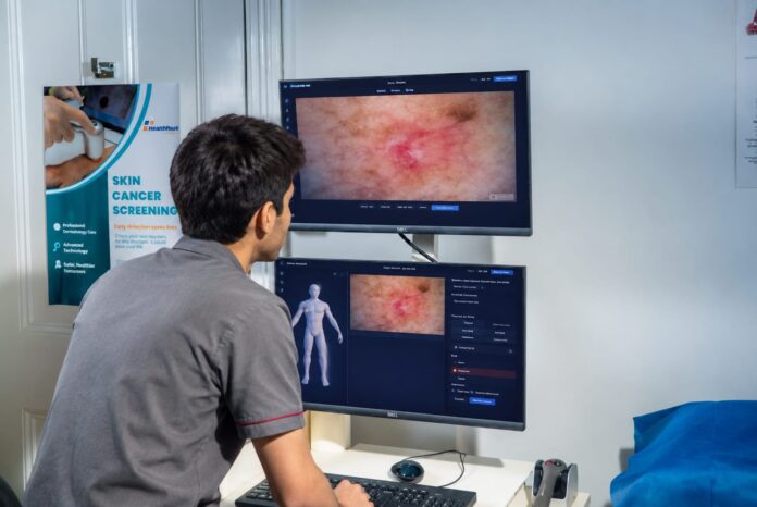
The gap between nakeds-eye accuracy and what proper tools can achieve is large and well documented. Handled dermoscopy, which examines lesions with magnification and specialized lighting that reveals structures below the surface, consistently outperforms the unaided eye. Pooled research found sensitivity for melanoma of about 90% with dermoscopy versus about 71% with nakeds-eye examination alone. Just as importantly, that improvement came with no decrease in specificity, meaning dermoscopy caught more cancers without wrongly flagging more harmless moles. That combination is rare and valuable. Usually, catching more disease means more false alarms, but here the accuracy simply improves.
For people at higher risk, such as those with many moles, a personal or family history of melanoma, or fair skin with significant sun exposure, a single examination is not enough, because the warning sign is so often change itself. This is where imaging over time becomes powerful. Modern digital mole-mapping systems let clinicians capture standardized, high-resolution images at each visit and compare a mole against its own documented history rather than a remembered impression. Total body photography has been shown to reduce unnecessary biopsies of benign moles and to help detect thinner melanomas than examination alone. In practice this means fewer people undergo procedures they never needed, while the genuine cancers are caught earlier.
What this means for you
If you are a patient, three habits meaningfully shift your odd.
- Know your own skin. Monthly self-checks using the ABCDE rule help you notice change early. A few phone photos taken periodically create a personal baseline you can compare against later, a low-tech version of what clinics do with imaging.
- Don’t talk yourself out of a worry. The cost of getting a harmless spot checked is trivial. The cost of ignoring a real one is not. Encouragingly, evidence from Melanoma Institute Australia suggests melanomas found during professional skin checks are associated with a lower mortality risk than those found by patients themselves. A trained eye and proper tools genuinely matter.
- Match your surveillance to your risk. If you have several risk factors, ask your clinician whether structured imaging and regular monitoring make sense for you, rather than relying on occasional glances.
If you are a clinician, the takeaway is blunts. Better tools translate directly into better-staged disease at diagnosis, and in melanoma, stage at diagnosis is destiny.
The bottom line
Melanoma is one of the few serious cancers where the single most powerful intervention is not a drug but a calendar. Finding it early is worth more than almost anything medicine can do once the disease has spread. The tools now exist to see earlier, to compare a mole against its own past, and to act sooner with confidence. Using them well, and encouraging patients to take changes on their skin seriously, is how a frightening word becomes, for the great majority of people, an entirely manageable diagnosis.
Sources
- Vestergaard ME et al. Dermoscopy compared with nakeds eye examination for the diagnosis of primary melanoma: a meta-analysis of studies performed in a clinical setting. British Journal of Dermatology, 2008.
- Kittler H et al. Diagnostic accuracy of dermoscopy. The Lancet Oncology, 2002.
- SEER Cancer Stat Facts: Melanoma of the Skin, National Cancer Institute.
- Systematic reviews on total body photography, Int. J. Environ. Res. Public Health, 2021.
- Melanoma Institute Australia, patient guidance.