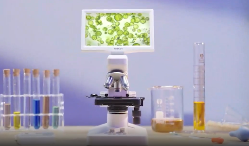

A single leaf conceals a bustling biological factory completely invisible to the naked eye. Exploring this miniature world reveals the intricate cellular structures powering plant life, from protective surface layers to microscopic breathing pores. Uncover the exact preparation techniques and optical settings needed to bring these botanical marvels into sharp focus.
What Does a Leaf Look Like Under a Microscope?
When you place a leaf under a lens, the solid green color instantly shatters into a complex, highly organized grid. Instead of a flat sheet, you see a three-dimensional landscape of interlocking geometrical blocks, snaking fluid tubes, and tiny moving green dots. It looks less like a simple piece of foliage and much more like a microscopic, densely populated city constructed from transparent bricks.
What Parts of a Leaf Can You Observe?
Because leaves are relatively thin, they are excellent subjects for transmitted light observation. With a properly prepared sample, several distinct biological layers and organs become immediately apparent.
Upper and Lower Epidermis
The epidermis is the outermost layer of the plant, functioning exactly like human skin. Under magnification, epidermal cells appear as a tightly packed, single layer of transparent puzzle pieces. They are highly structured and completely lack green color, acting as a clear protective window that allows sunlight to pass into the deeper tissues.
Chloroplasts Inside Leaf Cells
Beneath the clear epidermis lies the mesophyll layer, which contains the true engines of the plant. Inside these deeper cells, you will see dozens of vibrant green, oval-shaped structures called chloroplasts. These are the specialized organelles responsible for photosynthesis. In a fresh, living leaf, you can actually watch these green ovals slowly circulating around the edges of the cell, a process known as cytoplasmic streaming.
Leaf Hairs and Surface Structures
Many leaves feature specialized defense mechanisms on their exterior. Microscopic leaf hairs, known scientifically as trichomes, look like sharp, clear spikes or fuzzy, branched glass trees growing directly out of the epidermis. They protect the plant from hungry insects and help trap a layer of moisture against the leaf surface to prevent dehydration.
What Magnification Do You Need?
Plant cells are relatively large in the microscopic world, meaning you do not need extreme laboratory magnification to appreciate their beautiful structure.
Start at 40x for the Overall Structure
At 40x total magnification, a large portion of the leaf fills your field of view. While you will not see individual organelles clearly, this low power is perfect for observing the broader architecture. You can easily trace the thick, branching veins running across the tissue and locate dense clusters of spiky leaf hairs.
Use 100x to 400x for Leaf Cells
Increasing the magnification to between 100x and 400x brings the biological building blocks into sharp relief. At this range, the thick, rigid borders of the cell walls become distinctly visible. You can clearly differentiate the jigsaw-puzzle shapes of the epidermis from the rectangular cells stacked deeper inside the plant.
Increase Magnification for Stomata and Chloroplasts
To see the internal details of a single cell, 400x is the optimal setting. At this power, the tiny green chloroplasts become distinct, individual ovals rather than a blurry green wash. This is also the ideal magnification for finding and examining the precise crescent shape of the guard cells that form the plant’s breathing pores.
How to Prepare a Leaf for a Microscope
Dropping a whole, dry oak leaf onto a glass slide will just result in a dark, opaque blob. You must prepare the sample correctly so light can pass through the biological tissues.
Choose a Thin Fresh Leaf
The absolute best specimens for beginners are aquatic plants like Elodea, which are naturally only a few cell layers thick. If using a terrestrial plant, choose a fresh, soft leaf. Brittle, dried leaves trap dark air pockets and have completely degraded internal cellular structures.
Cut or Peel a Small Sample
You only need a fingernail-sized specimen. To see the surface cells, use precision tweezers to grip the very edge of a torn leaf and gently peel back a thin, transparent strip of the lower epidermis. To see the interior cross-section, use a sharp razor blade to slice the thinnest possible sliver across the leaf’s vein.
Place the Sample on a Slide
Lay your thin epidermal peel or cross-section perfectly flat in the center of a clean glass slide. Ensure the sample is not folded over on itself, as the overlapping cell walls will create a confusing, dense grid that is impossible to focus on clearly.
Add a Coverslip Carefully
Place a single drop of distilled water directly onto the leaf sample to keep the living cells hydrated and plump. Hold a glass coverslip at a 45-degree angle to the slide, touch the edge of the water drop, and let it fall gently over the sample. This angled approach pushes the air out and prevents frustrating microscopic bubbles from ruining your view.
How to Observe a Leaf Under a Microscope
Once your slide is prepared, navigating the biological landscape requires methodical optical adjustments.
Start With the Lowest Magnification
Always place your slide on the stage with the lowest power objective lens clicked into position. This provides the widest possible field of view, allowing you to easily locate the perfectly peeled section of the leaf before you attempt to look closely at the microscopic details.
Focus on the Leaf Surface
Use the large coarse focus knob to bring the surface of the leaf into view. You will know you have hit the correct focal plane when the sharp, rigid lines of the cell walls suddenly snap into clarity against the bright background.
Move to Higher Magnification
Center the most interesting part of the leaf—such as a cluster of stomata—perfectly in the middle of your light circle. Rotate the nosepiece to your higher magnification lens. Because high-power lenses sit incredibly close to the slide, only use your fine focus knob to dial in the final image sharpness.
Adjust Light for Better Detail
Plant cells are highly transparent, meaning too much bottom lighting will completely wash out their delicate boundaries. Closing your iris diaphragm slightly increases the contrast, making the transparent cell walls pop against the bright water medium. A system designed with integrated mechanical and lighting controls, such as the Tomlov DM301 Digital Microscope with X-Y Stage, highly streamlines this biological observation. The mechanical X-Y stage allows you to smoothly track moving chloroplasts without nudging the slide by hand, while the adjustable illumination helps you quickly dial in the perfect contrast on the large built-in monitor.
How to Identify Leaf Structures
Knowing exactly what biological feature you are looking at turns a simple observation into a true scientific exploration.
Find Cell Walls
Unlike round, squishy animal cells, plant cells feature a hard exterior shell made of cellulose. Under the microscope, these cell walls look like thick, dark, rigid lines separating the clear internal spaces. They form a continuous, highly organized grid that gives the leaf its structural physical strength.
Recognize Chloroplasts
Look inside the boundaries of the cell walls for distinct clusters of vivid green dots. These are the chloroplasts. Depending on the thickness of the leaf and the health of the plant, you may see anywhere from ten to over a hundred of these tiny photosynthetic engines packed inside a single cell.
Locate Stomata
Focus specifically on the lower epidermis (the underside) of the leaf. You are looking for structures that resemble tiny green coffee beans or microscopic mouths. These are the stomata, the physical pores the leaf uses to inhale carbon dioxide and exhale oxygen. The two crescent-shaped cells surrounding the opening are the guard cells, which swell and shrink to open and close the pore.
Identify Veins and Leaf Hairs
If you scan across the leaf, you will eventually encounter a thick, dark, fibrous bundle running linearly through the green cells. This is a vein, containing the xylem and phloem tubes that transport water and sugars. If you adjust your fine focus slightly upward, you can spot the sharp, translucent spikes of leaf hairs extending off the topmost layer of the epidermis.
Conclusions
Examining a leaf under a microscope bridges the gap between everyday nature and complex cellular biology. By learning to create a proper, thin wet mount and mastering your lighting controls on a WIFI digital microscope, you can strip away the plant’s solid green exterior to reveal the intricate, moving structures beneath. From the jigsaw puzzle of the epidermis to the actively photosynthesizing chloroplasts, this simple biological exercise offers a profound look at the tiny mechanisms that sustain life on Earth.